
436 N Roxbury Dr PH A, Beverly Hills, CA 90210
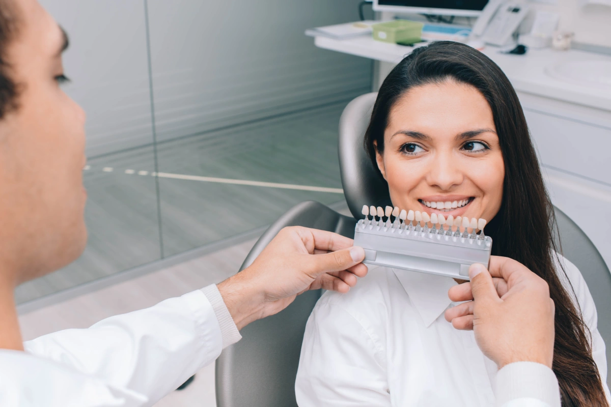
Porcelain veneers represent one of the most refined restorations in cosmetic and reconstructive dentistry. Crafted to exact specifications and bonded precisely to the tooth surface, they can deliver dramatic improvements in smile symmetry, color, and proportion. With proper care, they can serve patients well for many years. Yet even the most meticulously placed veneer can encounter problems — and when that happens, knowing why the emergency occurred is just as important as knowing where to turn.
Veneer emergencies in Beverly Hills are handled at the prosthodontic practice of Dr. Roxanne Salavati with the same level of attention and diagnostic precision that goes into creating the restorations in the first place. Understanding the root causes of these situations helps patients act quickly, avoid unnecessary delays, and protect the health of the teeth beneath.
What Counts as a Veneer Emergency?
A veneer emergency includes any situation in which a porcelain veneer chips, cracks, debonds, or begins causing discomfort that disrupts daily life. Some emergencies are immediate and unmistakable — a veneer that separates from the tooth mid-meal, for instance. Others unfold more gradually, beginning with mild sensitivity near the margin of the restoration or a faint darkening at the edge that slowly worsens.
Both types deserve prompt attention. What appears to be a surface-level inconvenience may actually reflect a condition developing within the tooth structure itself or at the interface between the veneer and the tooth.
The Four Most Common Causes of Veneer Emergencies
1. Decay Forming Beneath the Veneer
Porcelain does not decay. The natural tooth underneath it, however, can. When the seal between the veneer and the tooth is compromised in any way, bacteria can infiltrate the marginal area and establish a cavity in a location that is difficult to detect without advanced imaging.
The warning signs of decay beneath a veneer are easy to overlook because they mimic other conditions. Patients may experience sensitivity to cold temperatures or sweet foods, notice a darkened discoloration forming near the edge of the veneer, detect an unusual taste or odor, or find that the veneer begins to feel slightly loose. None of these signs should be attributed to normal wear without a proper clinical evaluation.
When caught early, subveneer decay can often be treated while preserving a significant portion of the remaining tooth structure. Waiting makes that outcome less likely.
2. Marginal Leakage
The margin is the boundary where the edge of the veneer meets the tooth surface. Even when this junction is sealed precisely at the time of placement, it can develop microscopic gaps over time. These openings — far too small to see or feel — allow saliva and oral bacteria to work their way beneath the restoration.
Once beneath the veneer, bacteria can cause decay, inflame the surrounding gum tissue, or irritate the dental pulp. The leakage itself typically results from one or more contributing factors: the natural aging of bonding resins over many years, cumulative wear from biting and chewing, heavy occlusal forces including bruxism, or minor surface damage to the veneer that creates a pathway for infiltration.
Marginal leakage rarely announces itself with dramatic symptoms. Regular monitoring at professional dental visits is the most reliable way to detect it before the underlying consequences become serious.
3. Bonding Failure
The bond between a porcelain veneer and the tooth is not created by the veneer itself — it depends entirely on the adhesive materials and technique used during cementation. Every step of that process is critical. Tooth preparation must be precise. The porcelain surface must be properly conditioned. Moisture must be controlled throughout. Cementation must follow exact protocols.
When any step is compromised — whether during the original placement or as adhesive materials age over many years — the bond can weaken. Moisture contamination during cementation, incomplete surface preparation of either the porcelain or the enamel, or breakdown of bonding resins over time can all result in a veneer that begins to loosen or eventually detaches.
The good news is that bonding failure does not always mean a veneer must be discarded. Many veneers that have separated from the tooth remain structurally intact and can be professionally cleaned, re-etched, and rebonded — provided that the underlying tooth is healthy and the veneer shows no significant damage.
4. Trauma and Chronic Bite Forces
A single impact — from a sports collision, an accidental fall, or biting into an unexpectedly hard object — can generate forces far beyond what porcelain veneers are designed to absorb. The result is often a chip, fracture, or full debonding of the restoration.
Chronic forces can be equally damaging, even when no single incident is identifiable. Patients who grind or clench their teeth, a condition called bruxism, place repeated high-magnitude stress on their veneers night after night. Over time, this can fatigue the porcelain, degrade the bonding interface, and ultimately lead to failure — even in veneers that were placed and maintained impeccably.
Evaluating the bite during every veneer emergency visit is essential. Without addressing the underlying occlusal issue, any repair or replacement is at elevated risk of failing again.
Pain Associated with a Veneer Requires Evaluation
Mild post-placement sensitivity is expected and typically resolves within days of veneer placement as the tooth acclimates. Persistent or newly developing pain is a different matter entirely. Discomfort originating near a veneer — whether a dull ache, sharp sensitivity, or pressure sensation — may indicate decay, pulp inflammation, a root infection, or a structural fracture extending below the gumline.
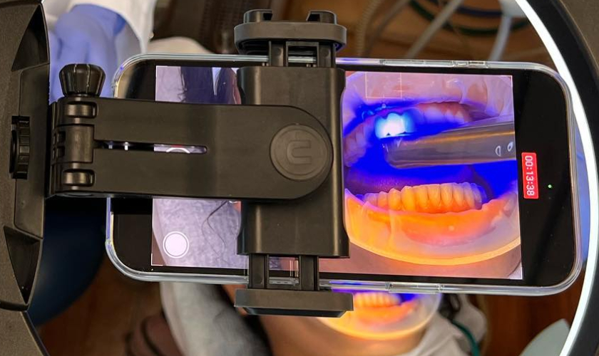
At Dr. Salavati’s office, veneer-related pain is evaluated with a thorough clinical examination supported by advanced imaging. Digital X-rays are obtained as an initial step. When the origin of the discomfort is not immediately clear or when deeper pathology is suspected, a Cone Beam CT (CBCT) scan is recommended.
CBCT technology produces a three-dimensional view of the tooth and its surrounding structures — something that two-dimensional radiographs cannot provide. With this level of imaging, Dr. Salavati can identify hidden decay beneath the veneer, cracks or fractures extending into the root, root infections, bone changes around the affected tooth, complications from prior endodontic treatment, and other conditions that would remain invisible on a standard dental X-ray. That diagnostic clarity makes it possible to recommend treatment that is appropriately targeted rather than presumptive.
Treatment Follows the Diagnosis
Because veneer emergencies arise from different causes, the correct treatment varies from patient to patient. After a comprehensive examination and accurate diagnosis, Dr. Salavati will recommend the course of care most appropriate for the specific situation. Depending on the findings, treatment may involve:
- Rebonding a veneer that has detached but remains intact and undamaged
- Replacing a veneer that has fractured, chipped significantly, or cannot be adequately rebonded
- Treating active decay beneath the restoration before fabricating a replacement veneer
- Root canal therapy when the dental pulp has become infected or irreversibly inflamed
- Replacing deteriorated bonding materials that have broken down over time
- Occlusal adjustment to redistribute bite forces and reduce stress on the restoration
- Fabricating a custom night guard for patients with bruxism or clenching habits
The overarching goal in every veneer emergency is to resolve the immediate problem, preserve as much healthy natural tooth structure as possible, and restore both the function and the appearance of the smile with lasting results.
Acting Early Matters More Than Most Patients Realize
It is tempting to take a wait-and-see approach when a veneer symptom seems minor. A veneer that rocks slightly but does not fall off. Sensitivity that is present but tolerable. A small discoloration at the margin that has not noticeably grown. These situations can feel manageable enough to defer — but the underlying processes causing them rarely pause.
Decay progresses into the tooth structure. Leakage allows bacteria to reach the pulp. What began as a rebondable veneer becomes a tooth requiring endodontic treatment. And what could have been a straightforward replacement becomes a more involved restorative case. Early evaluation does not just lead to simpler treatment — it consistently leads to better long-term outcomes for the tooth.
Veneer Emergencies in Beverly Hills: Specialized Care When It Matters
If a veneer has chipped, become loose, or begun causing discomfort, the first step is a thorough evaluation by a clinician with the training and diagnostic tools to identify exactly what is happening beneath the surface.
Dr. Roxanne Salavati is a prosthodontist in Beverly Hills with specialized training in complex restorations and the management of failing or damaged dental work. Prosthodontists complete three additional years of post-doctoral training beyond dental school, focusing exclusively on the restoration and replacement of teeth — a distinction recognized by the American College of Prosthodontists as the highest level of expertise in restorative care. With advanced diagnostic technology including CBCT imaging and a detailed understanding of the biology of tooth-veneer interfaces, her practice is equipped to identify the true cause of a veneer emergency and recommend treatment that addresses it precisely.
Patients experiencing veneer emergencies in Beverly Hills are encouraged to contact the office as soon as possible. Prompt evaluation protects the tooth, simplifies treatment, and gets the smile back on track.
Frequently Asked Questions
What should I do immediately if my veneer falls off?
Place the veneer in a clean container and contact a dental office as soon as possible — ideally one with same-day or next-day availability for emergency cases. Do not attempt to reattach the veneer using household adhesives or over-the-counter dental cements. These materials are not designed for bonding porcelain to tooth structure and can contaminate both surfaces, making proper professional rebonding more difficult or impossible. While waiting for your appointment, avoid chewing on the affected side and be cautious with temperature extremes, as the exposed tooth may be more sensitive than usual.
Can a veneer that fell off be put back on?
In many cases, yes — but only after a clinical evaluation confirms that rebonding is appropriate. If the veneer is structurally intact, the underlying tooth is free of decay, and the original debonding was due to adhesive failure rather than structural damage, a prosthodontist can often clean and recondition both surfaces and rebond the restoration successfully. If decay is present, the tooth surface is damaged, or the veneer itself has been compromised, replacement or additional treatment will be necessary before a new veneer can be placed.
Is it normal to have tooth sensitivity after getting veneers?
Some degree of sensitivity in the first few days following veneer placement is common and typically resolves on its own as the tooth adjusts to the restoration. What is not normal is sensitivity that persists beyond a week or two, returns after an initial resolution, or increases in intensity over time. These patterns suggest something beyond routine post-placement adjustment — possibly leakage, emerging decay, or pulp irritation — and should be evaluated promptly rather than monitored at home.
How can patients protect their veneers and lower the risk of emergencies?
Consistent oral hygiene, including brushing with a non-abrasive toothpaste and flossing carefully around the veneer margins, helps maintain the integrity of the seal. Regular professional cleanings and checkups allow a dentist to monitor the margins and detect early signs of leakage or wear. Patients should avoid using their teeth as tools, biting directly into very hard foods, or engaging in contact sports without a mouthguard. For those who grind or clench their teeth, wearing a custom-fitted night guard is one of the most effective measures available for extending veneer longevity.
What is a CBCT scan and how does it help with a veneer emergency diagnosis?
A Cone Beam CT (CBCT) scan is an imaging technology that captures a three-dimensional view of the teeth, roots, jawbone, and surrounding tissues in a single low-dose scan. Unlike conventional dental X-rays, which produce a flat two-dimensional image, a CBCT provides cross-sectional views from multiple angles, allowing a clinician to see structures that would be invisible on standard radiographs. In a veneer emergency, this level of detail is particularly valuable when pain has an unclear origin, when decay or a fracture is suspected to extend beneath the surface, or when prior root canal treatment may be involved. The result is a more accurate diagnosis and a treatment plan that addresses the actual cause of the problem rather than its surface presentation.